Technology
Imaging hepatocellular carcinoma using 89Zr-labeled anti-glypican-3 monoclonal antibody.
Immuno-PET, the tracking and quantification of mAbs using positron emission tomography (PET) cameras, is an exciting innovation to increase the comprehension of in vivo behavior and efficacy of mAbs. Immuno-PET has recently reached maturity in terms of technical development and is now entering the phase of broad-scale clinical application. This white paper will focus on immuno-PET using full-length mAbs and the associated application of the long-lived positron emitters zirconium- 89 and iodine-124.
New therapeutic mAbs already pose a heavy economic burden on national healthcare systems today. Recent national reports show that hospitals will not always treat all patients with the optimal mAb therapy due to excessive costs and limited likelihood of success. How can we improve the situation?
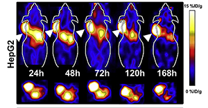
PET imaging of subcutaneous HepG2 cell xenografts with 89Zr -radiolabeled, anti-GPC3 monoclonal antibody clone 1G12.
Immuno-PET, the tracking and quantification of mAbs using positron emission tomography (PET), may offer a solution. It is a powerful innovation to improve the understanding of in vivo behavior and efficacy of mAbs. It combines the high sensitivity and resolution of a PET camera with the precision of a mAb.
Hepatocellular carcinoma is the third leading cause of cancer deaths worldwide. Since early detection may improve outcomes it is important to overcome the limitations of current imaging techniques such as ultrasound, computed tomography, and magnetic resonance imaging. These latter techniques are often incapable of detecting HCC lesions less than 2 cm and of differentiating between HCC lesions and other benign liver lesions leading to false positive results. Therefore, it is important to develop molecular imaging techniques which can improve the sensitivity and specificity of HCC detection.
Molecular imaging of cancer can potentially improve early diagnosis and clinical management. Positron emission tomography (PET) in using tumor targeting radio-labeled-molecules has gained wide acceptance in oncology, allowing improved diagnosis and clinical management of cancer patients. A variety of molecules including monoclonal antibodies (mAbs). antibody fragments, small proteins, peptides, and small molecules can be used as tumor-targeting molecules with different levels of tumor accessibility and specificity. The widespread availability of highly specific antibodies can lead to rapid advances in antibody-based probe development for PET imaging.
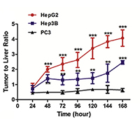 Tumor-to-Liver ratio of 89Zr -radiolabeled anti-GPC3 monoclonal antibody (clone 1G12) over time, for strong positive (HepG2), moderate positive (Hep3B) and negative (PC3) cell lines.
Generally, intact antibody molecules have relatively slow pharmacokinetics, which require multiple days to reach their optimal blood distribution within the body, therefore PET radioisotopes with the long half-life such as 89Zr (78.4 h) and (100.3 h). These differences are particularly suitable for intact antibodies, however since the liver is largely responsible for antibody clearance, the applicability of immuno-PET for HCC imaging remains unclear, with the major hurdle being high normal liver uptake and resulting poor tumor to liver ratio. The successful early detection of HCC lesions will require the combined selection of highly specific targets and effective approaches to decrease non-specific liver uptake of the imaging probe. Although other HCC associated biomarkers such as epidermal growth factor receptor (EGFR) has been used as an imaging target for HCC imaging, these approaches were typically associated with high liver backgrounds and unfavorable tumor-to-liver ratios.
In HCC, the heparin sulfate proteoglycan glypican-3 (GPC3) is a rational molecular target for HCC diagnostic imaging because it is: (i) a cell membrane receptor that is readily accessible for antibody mediated targeting and binding; (ii) expressed in more than 50% of HCC patients; (iii) capable of distinguishing malignant HCCs from normal liver, and pre-neoplastic and benign liver lesions; (iv) expressed at higher levels in small HCCs than in cirrhosis and other types of small focal lesions, suggesting that the transition from premalignant lesions to small HCC is usually associated with elevated GPC3 expression may aid early diagnosis.
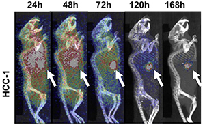
PET imaging of HCC xenografts derived from patient HCC with 89Zr -radiolabeled, anti- GPC3 mAb
It has therefore been hypothesized and that an immune-PET probe based on 89Zr -radiolabeled, anti- GPC3 mAb may offer the potential to accurately identify GPC3 positive HCC cells. This probe has been tested for its specificity for detecting HCC cells in vitro and in vivo and further determine its suitability for clinical translation using orthotopic HCC patient-derived xenografts.
GPC3 is an excellent molecular target in HCC, due to its preferential expression in HCC cells only, even in the early stages of malignant transformation. The intact monoclonal antibody (1G12) was used as the targeting molecule because of its demonstrated high affinity and specificity for recognizing GPC3. Given that an intact antibody has relatively slow pharmacokinetics, we rationalized that the use of along half-life radioisotope, 89Zr, might help to achieve clinically favorable tumor deliver ratios as it’s long decay half-life (3.3 days: 78.4 hrs) matches the biological half-life of the intact mAbs and thus, allows imaging at late times points (up to seven days) for obtaining maximum information. Additionally, the ability of 89Zr to be residualized and retained within the target cell after internalization and intracellular degradation of the tracer results in enhanced uptake in the tumor when an internalized antibody is used.
We have demonstrated that immuno-PET imaging of HCC based on GPC3 is a feasible and highly translatable approach. The specificity provided by the anti—GPC3 antibody, coupled with the high tumor-to-liver ratios provided by the 89Zr -mAb makes 89Zr-DFO-1G12 the most specific immuno-PET probe for HCC to date that can achieve high resolution imaging of GPC3-expressing HCCs.
Reference:
Imaging of hepatocellular carcinoma patient-derived xenografts using ⁸⁹Zr-labeled anti-glypican-3 monoclonal antibody.
Yang X, Liu H, Sun CK, Natarajan A, Hu X, Wang X, Allegretta M, Guttmann RD, Gambhir SS, Chua MS, Cheng Z, So SK. Biomaterials. 2014 Aug;35(25):6964-71.